Brian Cone Medical Group

BCMG AI

A Computer Vision Research & Development Company

Specializing in AI & Digital Pathology Tools, Whole Slide Imaging, Fluorescence Imaging & Metrology.

Locally Owned & Operated from Palos Verdes, CA — with Community & Educational Services at reduced or no cost.

Molecular Prediction · Gated ABMIL

ABMIL Research Platform

Gated attention-based multiple-instance learning on large WSI cohorts. All classifiers use a benchmarked panel of pathology foundation models -- including BCMG Emergent Encoding (in development). Ground truth from molecular profiling. No stain normalization applied. Benchmarked encoders: phikon-v2, UNI, and GigaPath.

Oncology · Thyroid
BRAF V600E — Papillary Thyroid Carcinoma
Algorithm Available
0.875
Best AUROC
Holdout, n=93
464
WSIs
96.1%
Peak Sens

Predicts BRAF V600E mutation from H&E morphology alone. Six-configuration benchmark, 3 encoders × 2 magnifications. Post-hoc QC pipeline: 130 slides excluded (28%), retrain achieved 47% variance reduction. NPV 93.3% at primary threshold.

EncoderAUROCSensSpec
UNI (40x)0.87588.2%76.2%
GigaPath (20x)0.84588.2%69.0%
phikon-v2 (40x)0.84296.1%69.0%
phikon-v2 (20x)0.84196.1%64.3%
UNI (20x)0.83394.1%69.0%
GigaPath (40x)0.83290.2%73.8%

Encoders: phikon-v2, UNI, GigaPath (pathology foundation models). No stain normalization.

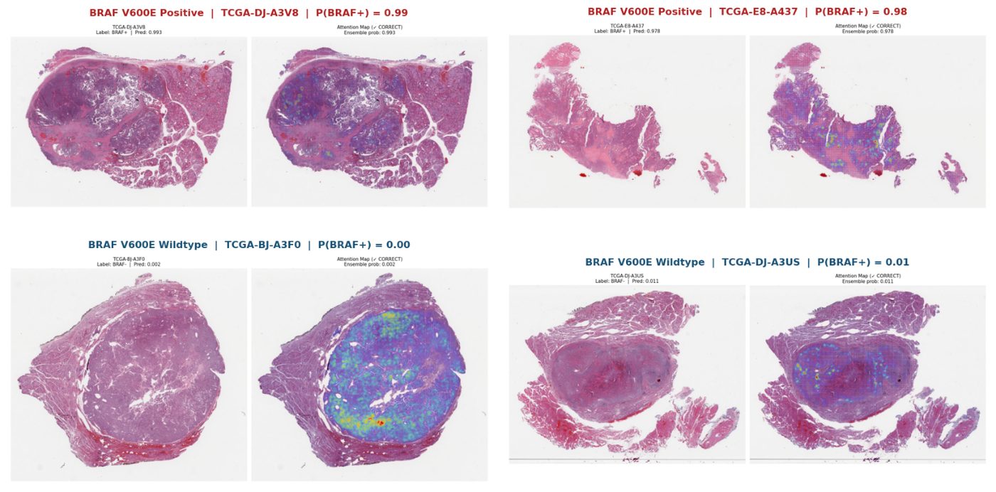
BRAF V600E attention heatmaps -- BRAF+ and wildtype cases

ABMIL attention heatmaps on four cases (two BRAF V600E+, two wildtype). Model probability shown per case. High-attention regions (warm colors) concentrate on tumor architecture.
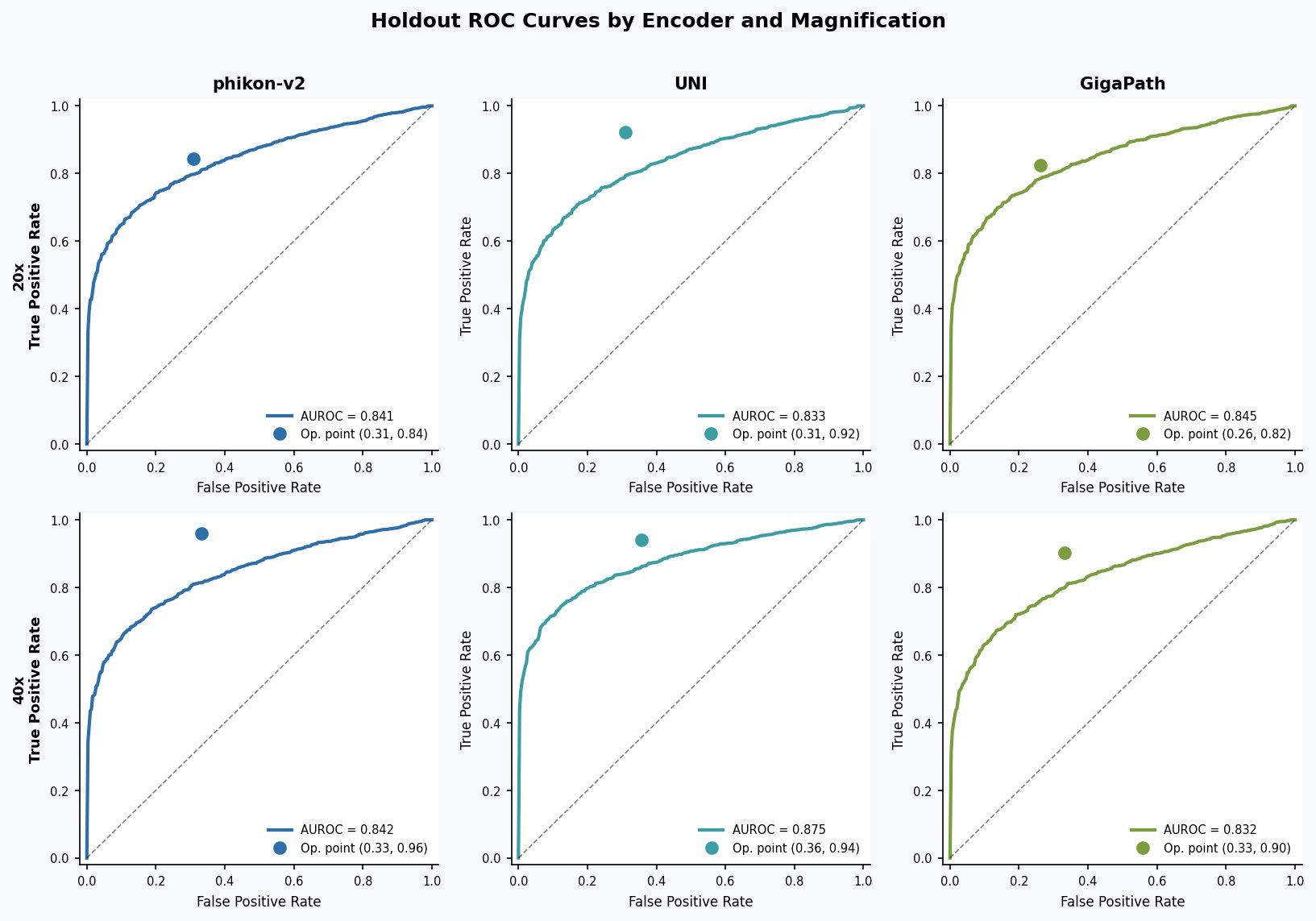
BRAF holdout ROC curves across 6 encoder-magnification configurations

Holdout ROC curves, all six encoder × magnification configurations. AUROC range 0.832–0.875.

Oncology · Colorectal
MSI-H Prediction — Colorectal Cancer
Complete
0.877
Best AUROC
Holdout, n=91
199
WSIs
92%
NPV
Rule-out threshold
Preprint coming License Algorithm

MSI-H vs. MSS from H&E. 14.3% prevalence -- substantially imbalanced. Rule-out threshold: 85% sens, 89% spec, 92% NPV, 65% screen-out. Three-encoder benchmark at 40x.

EncoderCV AUROCHoldoutSpec
GigaPath0.849 ± 0.060.87762.8%
phikon-v20.851 ± 0.100.83785.9%
UNI0.578 ± 0.080.60648.7%

Encoders: phikon-v2, UNI, GigaPath (pathology foundation models). No stain normalization.

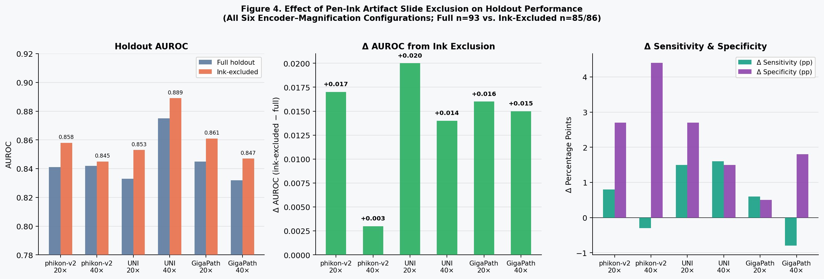
Effect of pen-ink QC filtering on holdout AUROC across encoder configurations

Effect of automated pen-ink QC exclusion on model performance. 28% slide exclusion, 47% CV AUROC variance reduction. Applied across all BCMG ABMIL pipelines as a pre-training gate.
Computational Pathology Kit (CPK): The BCMG pipeline packaged for on-premises deployment at academic medical centers. Partner institutions run the full ABMIL workflow on their own compute, against their own cohorts. Planned pilot: USC Keck Medical Center (collaboration with Dr. William Dean Wallace, Professor of Pathology).

Hematopathology · Multimodal
HemePath Aspirate Classifier
Active Development
0.989
Holdout AUROC
21-class marrow cells
82.6%
Accuracy
Held-out
0.909
Macro AUROC
AML subtype

Next-generation cellular segmentation and classification for the hardest hematologic specimens -- bone marrow aspirate smears and trephine, plus peripheral blood. The system is multimodal by design, fusing conventional smear morphology with raw event-level flow cytometry for a case-level differential -- a pairing not represented in the published literature.

Because aspirate smears are dirty, evaluation runs on spicule-containing, multilineage fields of view rather than whole slides indiscriminately -- the basis of BCMG hematologic precision scanning. Attention weights and Grad-CAM overlays make every slide-level call reviewable cell by cell.

TaskMetricValue
Bone marrow — 21 cell classesHoldout AUROC0.989
AML cytogenetic subtype (5-way)Macro AUROC0.909
NPM1 mutation — ABMIL on PB smearHoldout AUROC0.839
Plasma-cell detection (YOLO)mAP@500.85
Plasma-cell segmentationseg mAP@500.87

Research and decision-support stage; not a diagnostic device. Metrics are internal held-out results on public research cohorts. Contact info@bcmg.ai for methods.

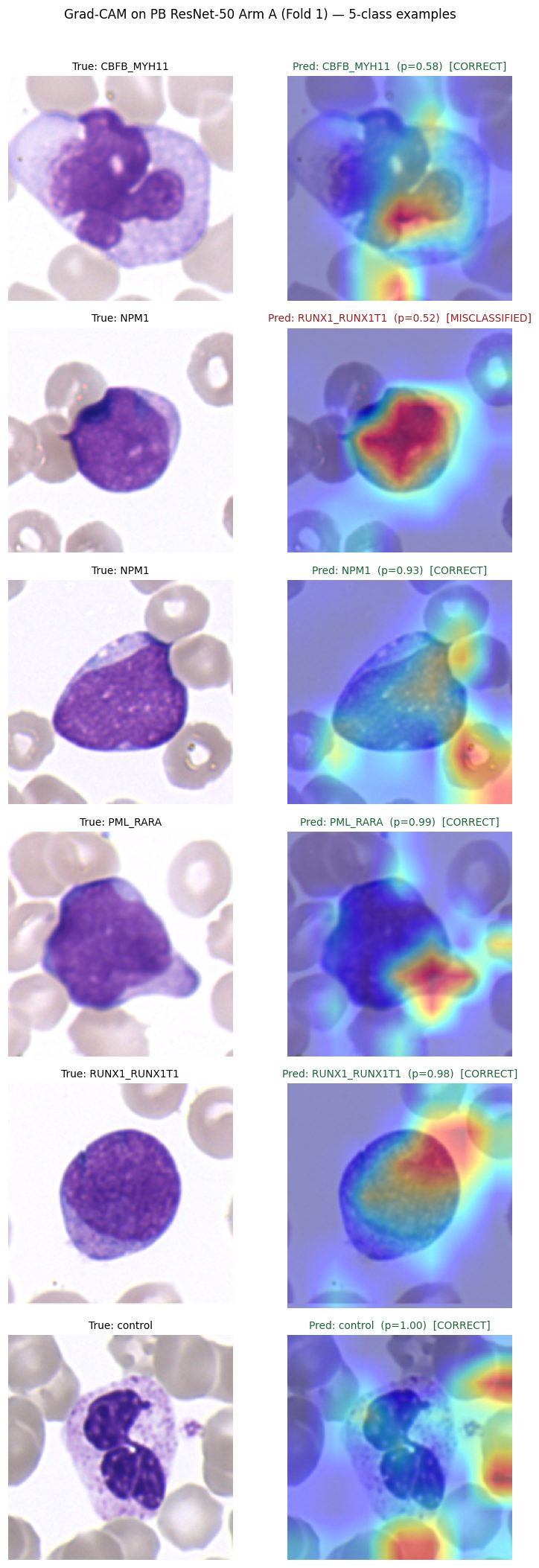
Grad-CAM attention overlays on peripheral-blood white cells across five AML cytogenetic subtypes

Peripheral-blood white-cell crops for five AML cytogenetic subtypes with Grad-CAM attention overlays and per-cell predicted labels. Network focus is inspectable per cell.
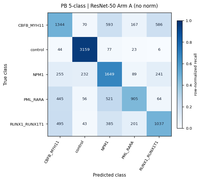
Row-normalized confusion matrix for the peripheral-blood AML subtype classifier

Row-normalized confusion matrix, peripheral-blood AML cytogenetic-subtype classifier.

On the roadmap: gated fusion of aspirate WSI features with a flow-cytometry set-encoder embedding into one calibrated differential, forward-compatible with 30+-marker spectral flow.

Cardiopulmonary
Pulmonary Hypertension H&E
Active — Preprint

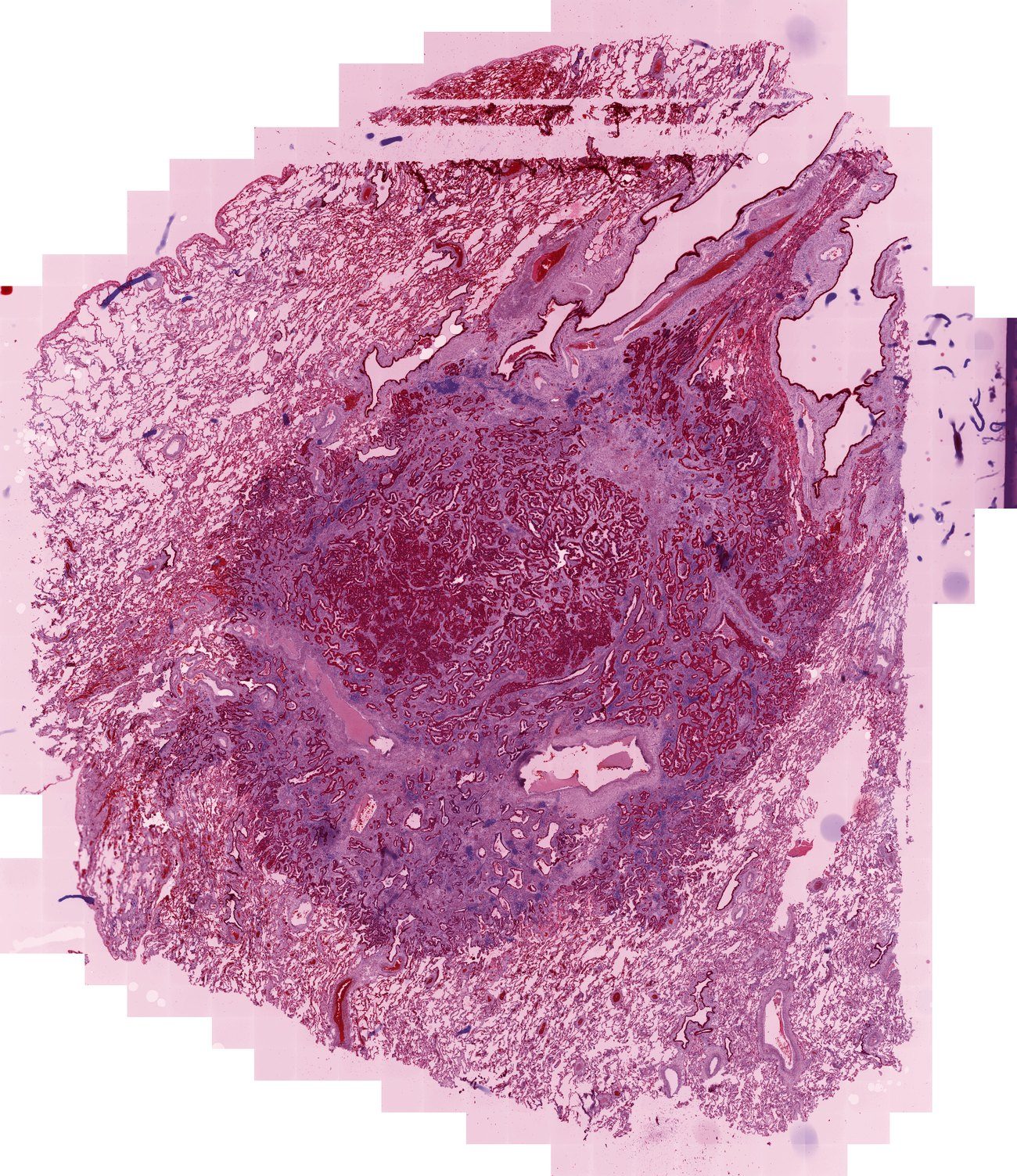
ABMIL classifier predicting pulmonary arterial hypertension from lung H&E. Collaborative study with Dr. William Dean Wallace (USC Keck): 18 explant cases, leave-one-out CV AUROC 0.975. Preprint available.

Oncology · NSCLC
MET Amplification — NSCLC
Queued

Slide-level MET genomic alteration detection from H&E. Early-stage; collaborators with relevant WSI cohort data are welcome to contact us.

Quantitative IHC · DAB2JETIA Platform

DAB2JETIA — Automated IHC H-scorer

A general anchored-colormap engine for quantitative DAB immunohistochemistry. Color stops map to the pathologist's own 0+/1+/2+/3+ scale -- not arbitrary per-slide histogram stretches -- so stain runs, slides, and cases stay directly comparable. TROP2 NSCLC below is the validation instance; the same engine extends to any DAB-developed biomarker and has spun out companion tools, including a counterstain-intensity scorer. Full method in the DAB2JETIA pamphlet.

Standard quantitative IHC heatmaps normalize DAB optical density to per-slide min/max. The cost: color no longer carries cross-slide meaning. A pixel rendered red on one slide may be 2+; on another, 3+. The semantics drift.

DAB2JETIA anchors the colormap on the standard semi-quantitative scale. OD ≤0.15 = 0+ (background) / OD 0.35 = 1+ / OD 0.70 = 2+ / OD ≥1.10 = 3+. A yellow pixel is definitionally between 1+ and 2+. A red pixel is at or above the 3+ anchor. Stain runs, slides, and cases become directly comparable.

Method: color deconvolution (Ruifrok H-DAB) isolates the DAB channel; piecewise-linear normalization maps optical density to pathologist-anchored stops. An optional multi-scale ridge filter emphasizes membranous structures without overriding cytoplasmic signal. Toggleable anchor-source architecture allows per-slide negative-control calibration with literature-default anchors as a non-destructive fallback. Architecture extends to HER2, PD-L1, and any DAB-developed IHC biomarker.
4
Scoring anchors
0+ 1+ 2+ 3+
5
Validation cases
TROP2 NSCLC
ICC > 0.87
vs Pathologist GT
TROP2 H-score
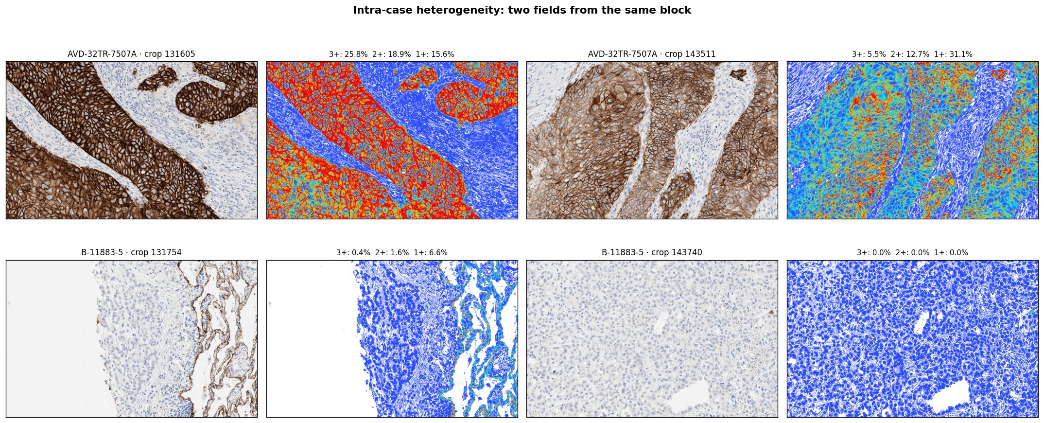
TROP2 IHC Scorer -- intra-case heterogeneity across two cases

Intra-case heterogeneity: two fields from the same block. Each pair shows original IHC and anchored heatmap with per-field 0+/1+/2+/3+ composition. Top: heterogeneous 3+ expression. Bottom: near-negative.
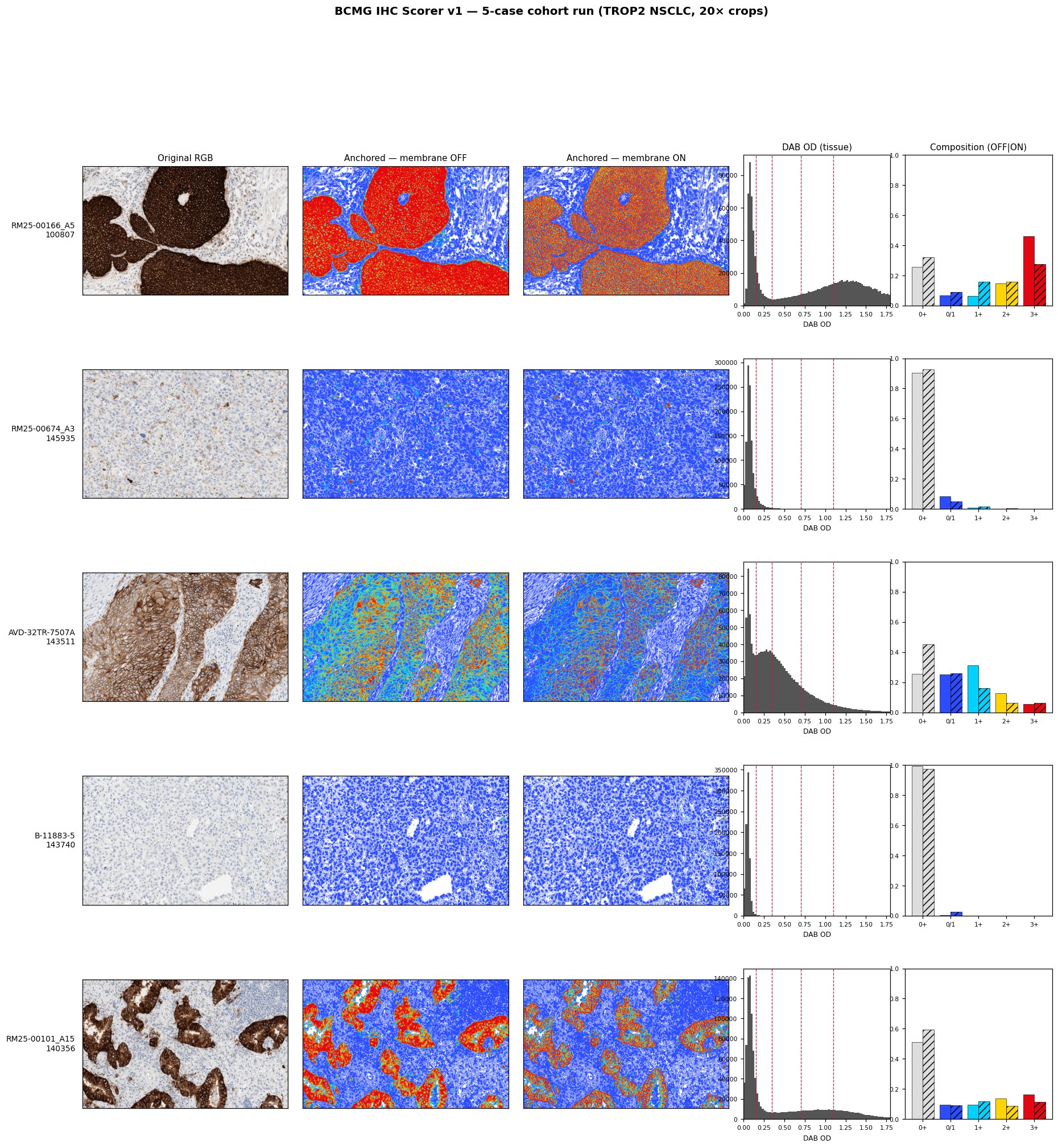
5-case TROP2 NSCLC cohort run -- full dynamic range

5-case cohort run (TROP2 NSCLC, 20× crops). Original RGB, anchored heatmap, DAB OD histogram, and composition bar per case. Full dynamic range from strong uniform 3+ to near-negative.
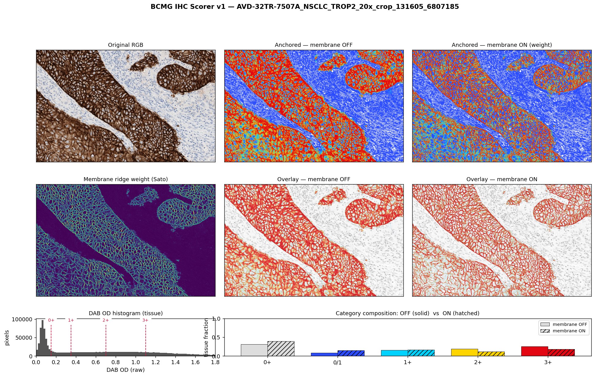

BCMG IHC Scorer -- single case detail panel with membrane ridge filter

Single case detail panel: original RGB, anchored heatmaps (membrane emphasis off / on), Sato ridge filter output, DAB overlays, and OD histogram. Membrane-emphasis layer toggles without overriding cytoplasmic signal.

Cloud-Native WSI Streaming

Nexus CloudView

Early Access

Open a gigapixel whole slide image directly from the cloud drive where it already lives -- Google Drive, OneDrive, Box, Dropbox, or Amazon S3 -- with no download, no copy, and no migration. To our knowledge, CloudView is the only viewer that streams a gigapixel WSI in place from consumer and enterprise cloud storage.

It works by HTTP range requests: the viewer reads only the byte ranges for the tiles currently on screen (FastAPI + OpenSeadragon DZI, OpenSlide-backed). A 1.9 GB slide opens in seconds because the rest of the file is never fetched. PHI stays inside your cloud tenant -- no BCMG server ever holds slide bytes.

  • Five storage backends: Google Drive, OneDrive, Box, Dropbox, Amazon S3
  • Reads Aperio SVS, Philips TIFF, and Hamamatsu NDPI in place
  • Only the tile bytes you view are transferred
  • QC annotation layer: Pass / Flag / Reject, CSV export
  • ABMIL attention-heatmap overlays
  • HIPAA-compatible: slide bytes stay in your tenant
CLOUD STORAGE CLOUDVIEW Google Drive OneDrive Box Dropbox Amazon S3 range requests visible tiles only only the lit tiles are fetched

CloudView reads only the byte ranges for the tiles in view. The rest of a multi-gigabyte slide is never transferred, and the file never leaves your cloud tenant.

Demo set: a curated CloudView demo spans multi-scanner format variety (the same tissue as SVS, NDPI, and TIFF), H&E across thyroid, colorectal, and kidney (ccRCC, pRCC, chRCC), and slides with known molecular ground truth (BRAF V600E, MSI-H) and deliberate artifact cases. Individual demo slides reach 155,175 × 95,102 pixels.

Real-Time DIY Acquisition

Nexus WSImager & Tesselerator

Stage 1 Prototype — May 2026

GPU-powered whole slide imager on a standard pathology scope. Core law: the live camera field-of-view is always the center of the screen. As the slide moves, captured tissue trails away and remains visible as part of the growing WSI -- 4K 60 fps in real time. First real-tissue WSI captured May 2026.

4K/60
fps capture
Elgato 4K Pro HDMI
32 GB
GPU VRAM
RTX 5090
<0.1ms
Focus/frame

Hardware: Nikon Eclipse 90i, Plan 40x / 0.65 NA, AmScope AF408 4K primary camera. Phase correlation + SIFT + optical flow registration. OME-TIFF output feeds CloudView and the ABMIL pipeline without conversion. Nexus-Nikon Stage Mapper runs in parallel for motorized ASI XY acquisition.

Why it matters: a clinically useful WSI without a $200,000 scanner -- built live, by hand, on the microscope a lab already owns.

Tesselerator calibration -- 2x scan tissue mapper on the BCMG AI fiducial print slide (edited to key motion).

Nexus WSImager -- 2x FullView WSI companion demo.


Quality Control

Digital Pathology QC Tools

Pen Ink Artifact Filter

A research-pipeline-integrity gate for deep and agentic computer-vision pathology. Pen and surgical inks are non-biological, spectrally extreme pigments; left in the tile bag, an attention model latches onto them and learns artifact instead of morphology. The filter intercepts ink tiles before ABMIL ingestion, so the model is not silently trained on corrupted signal.

Two-track automated pipeline. Track A: HSV color + MLP for speed. Track B: slide-level ABMIL on pathology features for robustness. Validated on a 464-slide thyroid cohort: 28% exclusion rate, 47% AUROC variance reduction downstream. Runs as a pre-training QC gate on all BCMG pipelines.

NEXUS HTML Viewer

Multi-source document viewer and version comparer for pathology reports and research HTML artifacts. 147 documents indexed across 13 project categories. Full-text search, project/type/owner filtering, inline annotation, side-by-side diff comparison, PDF export. Standalone -- no server required. Available for evaluation on request.

Nexus Imaging System

Full integration dashboard for the Nikon Eclipse 90i: objective inventory (nine lenses, 2x–50x oil), per-objective Abbe resolution and depth-of-field calculations, multi-camera module registry (AmScope AF408 4K, ELP USB-3, ArduCam MIPI), and stage/focus configuration. Built for the WSImager and Tesselerator workflows.


Scanner Normalization

HistoLux

Active Design

Scanner-agnostic color, gamma, and white-balance normalization for histopathology WSI. Scanners differ in gamma, white point, and effective color space -- a Hamamatsu NanoZoomer and a Leica AT2 render the same section differently, shifting the input distribution for models trained on Leica-dominant public data. HistoLux corrects this at the image level before features are extracted.

A four-stage, physics-principled pipeline: background/glass detection, gamma calibration, Von Kries chromatic adaptation, and Macenko stain-density matching -- interpretable, with no training data required.

Reference standard: Leica AT2 / GT450 (dominant in TCGA). Correction targets: Hamamatsu NanoZoomer S60 / S360. Scanner profiles are stored as JSON and applied once characterized; validation targets SSIM > 0.85 and a positive AUROC delta on corrected vs raw slides.

Slide Labeling & Provenance

C4D Label System

In Development

A color 2D barcode slide label. Beyond an immutable slide ID, the label carries a scan-request matrix -- four color positions that encode exactly which acquisitions a slide should get: a 2x or 4x full-slide companion, and 20x or 40x WSI. The physical label itself tells the imaging system what to do.

It is built to route a slide through BCMG's own imaging (Tesselerator and Nexus WSImager) and to travel with the slide for external WSI marketplacing, for example Biobase. Because the label records what was requested, a lab keeps a clean separation between requested work and the actual acquisition metadata the imaging system later records -- useful whether or not the lab works with BCMG.
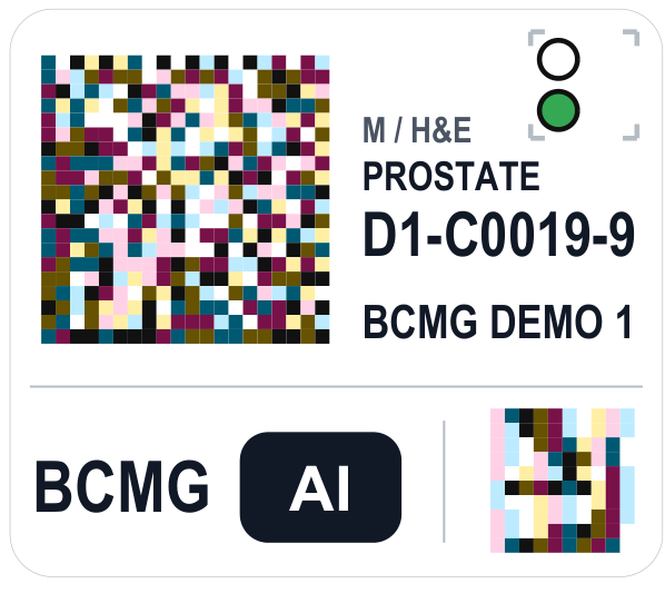
BCMG AI C4D color 2D barcode slide label with scan-request matrix

A C4D label: immutable slide ID, human-readable specimen line, and the four-position scan-request matrix (white 2x / red 4x companion, green 20x / blue 40x WSI).

Bridges imaging and marketplace: the same label routes a slide through BCMG acquisition and carries a partner-friendly identity for external supply. See BCMG Research Tissues for the marketplace side.
Clinical Computer Vision · Wound Metrology

Wound Tracker & the 3-D Wand

Ruler-free wound measurement from an ordinary image -- calibrated area, dimensions, tissue composition, and a registered map of change over time. Proprietary BCMG metrology does the measuring, the clinician reviews every result, and nothing leaves the clinic.

From Image to Measurement

Objective Measurement & Tracking

In Development

Wound care runs on measurements, yet everyday documentation still leans on paper rulers, tracings, and visual estimates that vary by observer and visit. BCMG AI turns a simple photograph -- with a single-use printed reference tag beside the wound to set absolute scale -- into a reproducible measurement: area, greatest dimension and perpendicular width in millimeters, wound-bed tissue breakdown, and a visit-to-visit change map.

A promptable vision model delineates the wound. The clinician stays in control: any boundary can be accepted, rejected, or corrected with a single click-and-drag, and any result outside strict quality bounds is flagged rather than silently reported.

  • Area, greatest dimension, perpendicular width -- in millimeters
  • Wound-bed tissue composition: necrotic / granulation / epithelial
  • Registered change map -- every point colored by how the margin moved
  • Serial trend across visits, with automatic resolution detection
  • On-prem and HIPAA-compatible -- images and PHI stay in the clinic
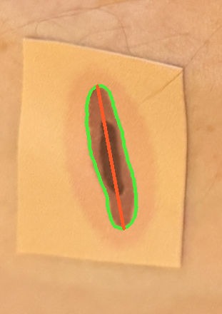
Automated wound delineation: green boundary and red greatest-dimension on a scale-rectified frame

Automated delineation: wound boundary (green) and greatest dimension (red) on the scale-rectified frame. Development test wound.


Handheld Capture · Calibrated Stereo

The BCMG AI Wand

Active Design

A handheld, factory-calibrated stereo imaging device that standardizes capture in the clinic. Because its two matched cameras are stereo-calibrated, absolute scale comes from the 3-D geometry itself -- so nothing is placed near the wound. Its job is consistency: the same framing and working distance at every visit, so serial measurements track the wound rather than the photographer.

On-screen guidance keeps the operator inside the validated working band and warns when lighting or texture would degrade a measurement -- capture problems are caught while capturing, not after the patient has left. Where the surface allows, the same 3-D geometry yields wound depth and volume that a flat photograph cannot provide.

  • No fiducial on the skin -- calibrated stereo recovers scale directly
  • Standardized distance and framing at every visit
  • Any trained operator: follow the reticle, sweep, done
  • Depth and volume where surface shape allows
  • Closed-loop, on-prem device and workstation
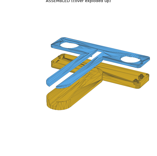
BCMG AI Wand stereo module and optical cover, development CAD exploded view

The Wand's stereo module and optical cover -- development CAD, exploded view.
Human-in-the-loop, on-premises: every automated result is the clinician's to accept, adjust, or reject, and the full workflow -- capture, measurement, storage -- runs inside the clinic. In active development; figures show development test wounds and CAD. Research and evaluation inquiries: info@bcmg.ai.
Primary Commercial Product · Palos Verdes Peninsula

BCMG Movement Index

Per-parcel 0–100 ground displacement score. Buyers, sellers, real estate professionals, and risk underwriters get a legible, scientifically grounded signal about whether the ground under a parcel is moving -- and how fast -- based on millimeter-precision satellite radar measurements.

86K+
Valid pixels
PV Peninsula
38.5
mm/yr peak
2016–2024 baseline
16
Markets live
5 continents

Built on ALOS-2 PALSAR-2 L-band InSAR (JAXA). L-band (23 cm wavelength) penetrates vegetation canopy and maintains coherence in coastal fog -- where C-band alternatives decohere and fail. All ascending and descending track pairs are composited into a single MI layer; single-track LOS velocity is never exposed as a consumer deliverable.

The Portuguese Bend landslide complex has measured movement from <0.1 mm/yr in stable zones to >300 cm/yr during acceleration events. Properties in the zone routinely transact above $2–5M. The BCMG MI is the first per-parcel InSAR intelligence product for this market.

Processing: Sentinel-1 SBAS pipeline (MintPy 1.6.3) established the methodology; ALOS-2 PALSAR-2 SLC processing is the primary production route. AI pipeline (Son of Satori) layers InSAR time-series with optical imagery, terrain slope, soil composition, and historical event data.

Join the waitlist for parcel-level scores in your area.

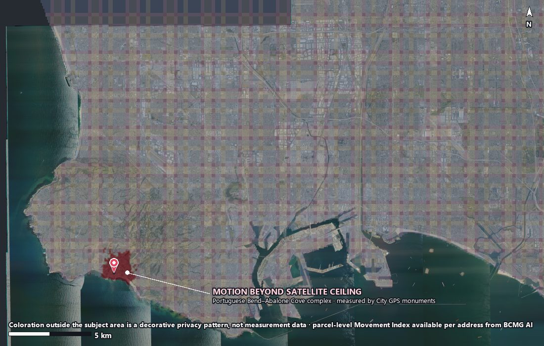
BCMG Movement Index peninsula overview -- Palos Verdes, ALOS-2 InSAR

BCMG Movement Index over the Palos Verdes Peninsula. The Portuguese Bend-Abalone Cove complex moves beyond the satellite ceiling; per-parcel scores are available by address. Coloration outside the study area is a privacy pattern, not measurement data.


In the Field · Palos Verdes

Why we validate on Portuguese Bend

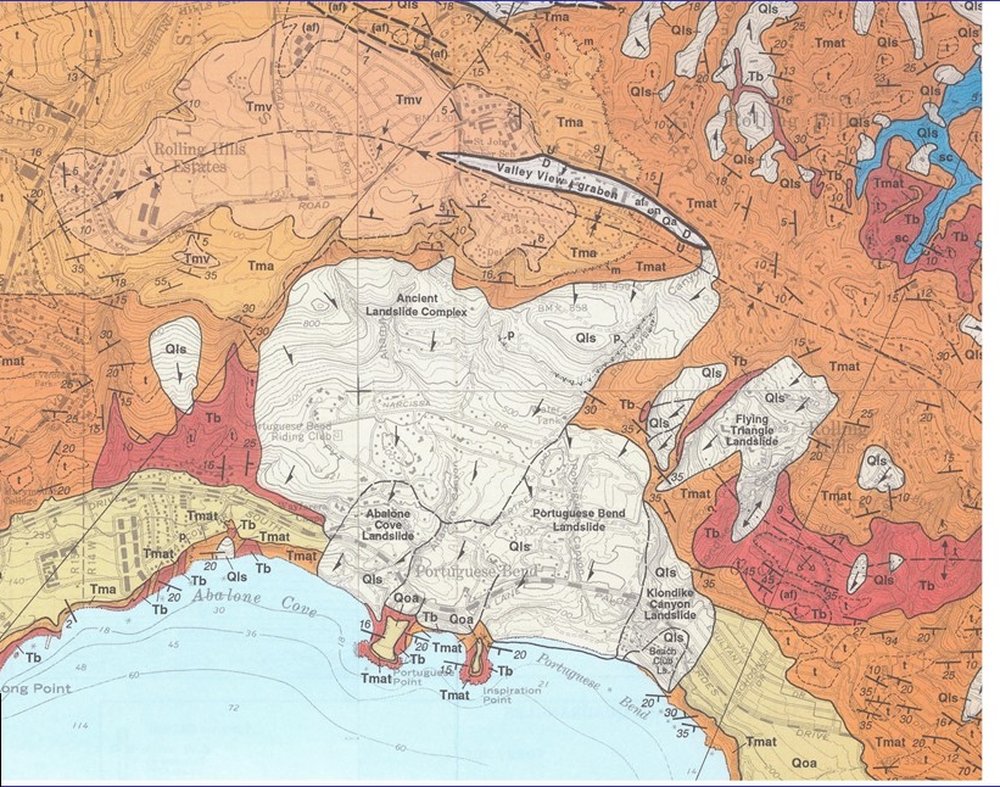
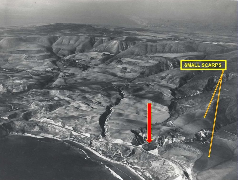
The Portuguese Bend landslide complex is one of the most extensively studied active landslide systems in the United States -- with well-documented movement since the mid-20th century and the landslide terrain recorded in aerial surveys as early as 1931. That deep, public record is exactly why we validate here: ground truth is abundant, and the fastest-moving zones push the very limits of what satellite radar can measure.

Two points matter for reading any Movement Index map. First, the Index is not specific to Portuguese Bend, or to Palos Verdes -- the same per-parcel scoring runs anywhere Sentinel-1 or ALOS-2 coverage exists, from a single hillside lot to a metropolitan submarket. Second, an active complex sits inside a peninsula that is overwhelmingly stable: the great majority of parcels read green. The value of the Index is precisely that separation -- telling the specific ground that is moving from the far larger area that is not, with objective per-parcel evidence rather than a blanket label.

The complex is not only historically documented -- it is actively measured. The City of Rancho Palos Verdes runs a formal Landslide Management Program, and annual GNSS (satellite-GPS) surveys have tracked the ground monument-by-monument since 2007. Recent surveys resolve movement against a noise floor near 0.02 ft, with the most active monuments in the Abalone Cove and Klondike Canyon slides projecting on the order of two feet per year -- while the surrounding ground holds still. That is exactly the regime a Movement Index is built to read, and exactly why the fastest cores can decorrelate satellite radar even as ground GPS still resolves them.

Geologic map of the Portuguese Bend / Altamira landslide complex, Palos Verdes

The mapped complex -- Portuguese Bend, Abalone Cove, Klondike Canyon, and Flying Triangle slides within the ancient Altamira landslide. Source: City of Rancho Palos Verdes.

1931 aerial photograph of the Klondike and Beach Club landslide area on the Palos Verdes coast

Surface features in a 1931 coastal aerial of the Klondike / Beach Club area, small scarps annotated. Source: City of Rancho Palos Verdes.


Agentic Pipeline · Human-Audited

How the Index is built

The Movement Index is not raw radar. An agentic workflow integrates InSAR displacement time-series with optical satellite imagery, terrain slope, soil composition, and historical event data into a single per-parcel score.

A supervised, human-in-the-loop step keeps it honest -- the same audited-review discipline BCMG brings to pathology. Candidate sites surfaced by heuristic and model scoring are reviewed and labeled by an expert against a fixed schema, and that feedback trains and validates the optical-evidence detectors. Optical review runs on NAIP aerial imagery.

Candidate-review schema

Collapsed / Damaged Landslide scar Debris Construction Vegetation / False+ Unsure

Each candidate chip of optical imagery is reviewed and labeled by an expert; those labels feed back into the detector -- a pathologist's audited-review loop applied to satellite evidence.


Next Generation · ALOS-2 L-band

ALOS-2 L-band portal (experimental preview)

The production Movement Index is moving to ALOS-2 PALSAR-2 L-band, whose long wavelength holds coherence over the fast-moving Portuguese Bend core where C-band decorrelates. This preview shows single interferometric pairs (ISCE2) over the peninsula: line-of-sight displacement, rewrapped phase fringes, and coherence.

Experimental, not validated -- internal review render only. Single pairs, no velocities, not yet a Movement Index.

ALOS-2 L-band single-pair interferometry over the PV Peninsula (ISCE2). Experimental preview.

InSAR · SBAS · 16 Active Markets

Global Markets & Conflict Spatial Analysis

US Domestic Subsidence

Sentinel-1 SBAS coverage across major US subsidence markets: LA Metro Basin (ASC+DSC composite, 2026-04-20), Houston (industrial subsidence, high-magnitude), Miami / Orlando / Hollywood (karst and coastal compaction), Santa Barbara, and Hawaii (volcanic deformation). All tiled and live in the NCV Geo Edition viewer.

For oil and gas operators, city planning departments, and insurance underwriters, BCMG provides custom InSAR analysis, per-parcel MI scores, and integrated risk layers for specific geographic areas of interest. Contact for scope and pricing.

Conflict Spatial Analysis

Pre- and post-event SBAS baselines combined with HyP3 rapid-response displacement products and PWTT conflict damage layers -- derived from the Post-War Terrain Transition algorithm developed by Ollie Ballinger (UCL, Remote Sensing of Environment, 2025). BCMG uses PWTT damage probability maps as an independent ground-truth validation layer for MI signals over the same regions.

Active coverage spans Middle East and Eastern Mediterranean post-conflict zones, Central Asian strategic sites (nuclear and industrial infrastructure deformation monitoring, 5–10 year chronic records), coastal and maritime chokepoints, and a labeled border-zone dataset supporting a tunnel detection CNN research program.

Applications: structural risk modeling for insurers, humanitarian damage assessment, academic collaboration, and geospatial intelligence research. All analysis uses open and commercial satellite data sources exclusively.


International · Coming Soon

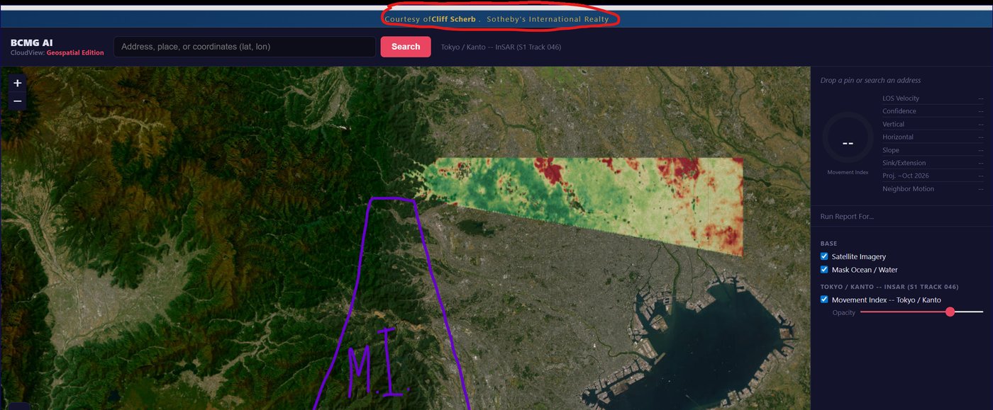
Tokyo / South Kanto

The Movement Index platform is not limited to Palos Verdes. A North Tokyo wedge of Movement Index data is already in the pipeline, extending BCMG's per-parcel InSAR intelligence to the South Kanto market. More to follow.

BCMG Movement Index -- North Tokyo InSAR wedge over the South Kanto region

CloudView Geospatial Edition: a North Tokyo Movement Index wedge (Sentinel-1 InSAR) over South Kanto. Sandboxed submarket preview -- warm tones flag higher movement, green reads stable.


Special Research Report · Prepared for NIST

Champlain Towers South — Surfside, Miami

Under Investigation

On 24 June 2021 the twelve-story Champlain Towers South condominium partially collapsed, killing 98 -- one of the deadliest peacetime building failures in US history. In June 2026 the NIST investigation attributed the initiating event to punching-shear failure at pool-deck and garage column-slab connections, beginning roughly three weeks before the collapse; ground subsidence was not a primary driver.

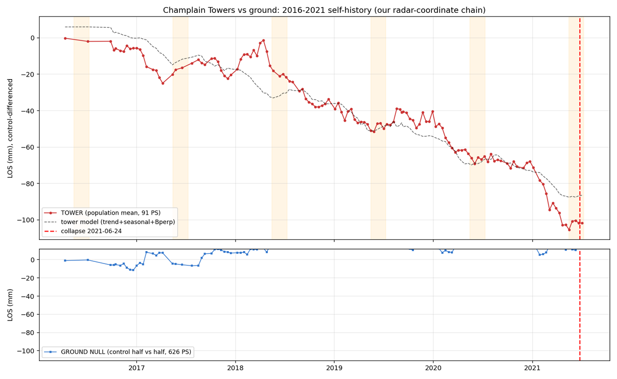
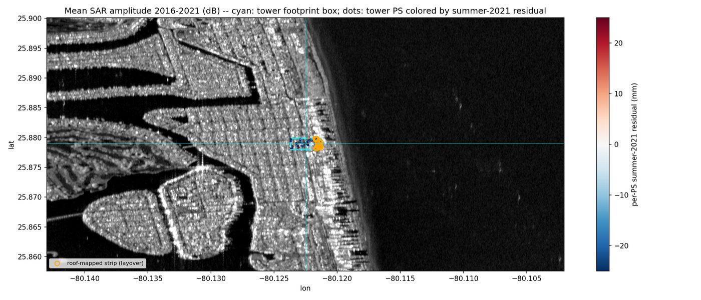
BCMG has prepared a special InSAR report for submission to NIST. Its central finding is stated as a disciplined null: a side-looking satellite cannot image the at-grade garage where the failure initiated, so InSAR could not have predicted this specific structural failure. A persistent-scatterer re-analysis (OPERA CSLC-S1, 134 acquisitions 2016–2021) finds the tower footprint is not a differential-subsidence hotspot. A summer-2021 line-of-sight excursion of −14.9 mm (z = −2.67) is the largest deviation from the tower's six-year average, but sits near the C-band noise floor and is held as suggestive, not conclusive.

The operative distinction for NIST: InSAR is a risk-zoning instrument, not a structural-failure predictor. It could have flagged the building as sitting in an active regional coastal-subsidence corridor warranting inspection -- consistent with Aziz Zanjani et al. (2024), which mapped up to 8 cm of subsidence across nearby Sunny Isles Beach.

Recommendations to NIST: mandate retrospective InSAR review in coastal structural-failure investigations; fold subsidence screening into high-rise recertification; commission a post-hoc PS-InSAR facade study; and keep risk-zoning distinct from failure prediction.

InSAR time series: tower persistent-scatterer displacement vs stable ground, 2016 to 2021

Persistent-scatterer displacement of the tower vs stable ground, 2016–2021. The summer-2021 excursion is visible but near the radar noise floor.

Annotated Sentinel-1 SAR amplitude scene over Surfside with the tower footprint marked

Annotated Sentinel-1 SAR scene over Surfside; the Champlain Towers footprint and land/ocean edge are marked.

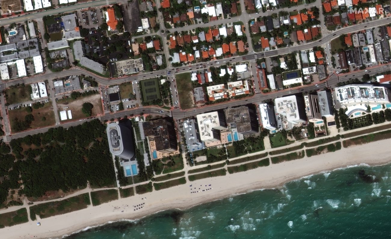
Maxar satellite image of Champlain Towers South intact before the collapse

Maxar optical, before -- the intact tower.

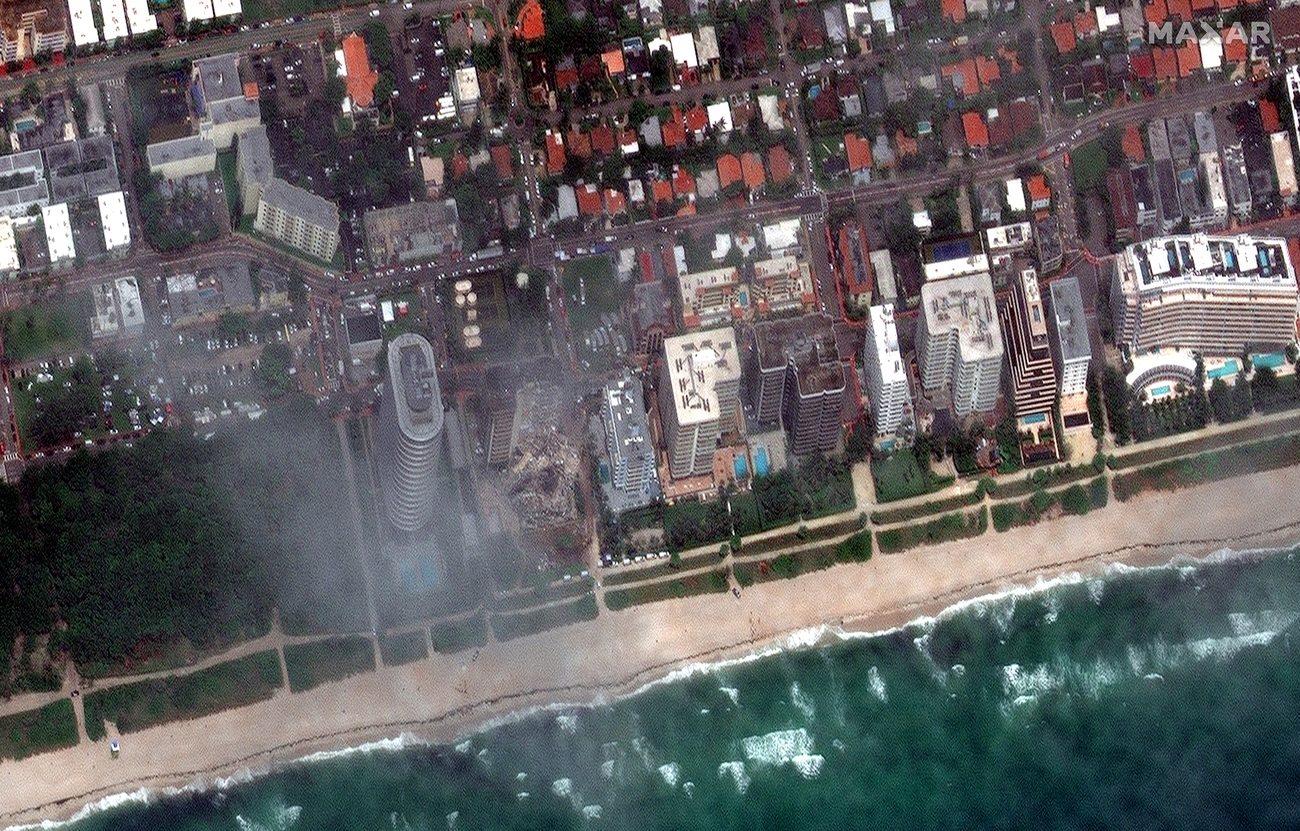
Maxar satellite image after the June 2021 collapse showing the debris field

Maxar optical, after -- the debris field, 25 June 2021.

Analysis uses open and commercial satellite data only. InSAR findings are stated as risk-zoning context and a methodological null, not a claim of a predictable structural precursor.

Joint Venture · Computational Pathology

Computational Pathology Kit

A joint venture between BCMG and Dr. William Dean Wallace, Professor of Pathology at USC Keck School of Medicine. CPK packages the full BCMG ABMIL pipeline for on-premises deployment -- giving academic pathology departments the computational capacity they need without requiring an in-house computational pathologist.

The Problem CPK Solves

Most pathology departments have zero computational pathologists. The field is moving fast -- biomarker prediction, IHC quantification, ABMIL-based molecular classification -- and departments without in-house expertise are being left behind.

CPK is a productized version of the BCMG research platform: slide ingestion, tiling, QC, feature extraction, attention-based inference, and report generation. Everything needed to run a production-grade ABMIL workflow on local hardware, with no cloud dependency and no data leaving the institution.

The planned pilot deployment is at USC Keck Medical Center, in collaboration with Dr. Wallace's pathology group.

0
Cloud Dependency
On-Prem
Deployment
ABMIL
Core Engine

Dr. William Dean Wallace — USC Keck

Dr. Wallace is Professor of Pathology at the Keck School of Medicine, University of Southern California. He brings deep domain expertise in pulmonary, cardiopulmonary, and renal pathology, institutional deployment experience, and an established research program in digital pathology.

Current collaboration: ABMIL classifier for Pulmonary Arterial Hypertension prediction from H&E whole slide images. Results under review for publication.

Cardiopulmonary · USC Keck Collaboration
Pulmonary Hypertension H&E Classifier
Active — Pending Publication

ABMIL slide-level prediction of Pulmonary Arterial Hypertension from lung H&E morphology. Collaborative study: Dr. Cone (BCMG) & Dr. Wallace (USC Keck). Results under review.


Interested in CPK?

CPK is currently in pre-release, with pilot deployment planned at USC Keck. Academic medical centers, health systems, and reference laboratories interested in bringing ABMIL-based computational pathology on-premises are welcome to reach out.

We respond to substantive inquiries within 48 hours. NDA available on request prior to technical disclosure.

Current Status: Pre-release. Pipeline validated on TCGA-THCA (BRAF V600E, n=464) and TCGA-COAD/READ (MSI, n=199). On-premises packaging and institutional license terms in development. Pilot site: USC Keck Medical Center.
Forward-Looking · Pharma Partnerships

Drug Discovery & Companion Diagnostics

An emerging direction for BCMG: applying quantitative IHC and attention-based whole-slide models to pharma R&D and companion-diagnostic development. This is a program we are actively developing, not a shipped product -- but each approach below is grounded in a tool BCMG has already built.

In Development — Partnerships Welcome
Approach 1
Quantitative IHC → sharper CDx
BCMG's anchored DAB quantification (the TROP2 IHC Scorer, DAB2JET lineage) turns a companion-diagnostic stain from an eyeballed 0/1+/2+/3+ call into a continuous, reproducible, cross-slide-comparable score (ICC > 0.87 vs pathologist H-score). A machine-anchored readout tightens the patient-selection cutoffs that antibody-drug-conjugate and targeted-therapy CDx depend on.
Approach 2
ABMIL for target discovery
The same gated attention-based MIL that predicts BRAF, MSI, and NPM1 status from H&E can be inverted: attention maps localize the morphology driving a molecular label, surfacing candidate tissue phenotypes and spatial niches tied to a target or a response. Trained across responder / non-responder cohorts, attention becomes a hypothesis generator for mechanism and stratification biomarkers.
Approach 3
Multiplex + virtual H&E readouts
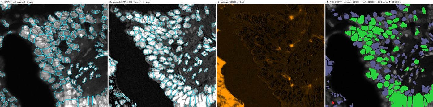
BCMG's mIF-to-pseudo-H&E and CD68-recovery pipelines let a single multiplex acquisition yield both a morphologic read and quantified immune-microenvironment features (tumor-macrophage fraction, spatial phenotyping) -- a pharmacodynamic and immune-context readout for trials, from one slide, without cutting extra sections.

Where this is grounded

These are not speculative capabilities. The quantitative IHC scorer, the ABMIL classifiers (BRAF AUROC 0.875, MSI 0.877, NPM1 0.839), and the multiplex and pseudo-H&E tools are all built and documented elsewhere on this site. The step to pharma is applying them to sponsor cohorts under the right agreements.

BCMG operates under NDA and can serve as a computational partner to biotech and pharma teams developing tissue-based biomarkers, companion diagnostics, and response-stratification models.

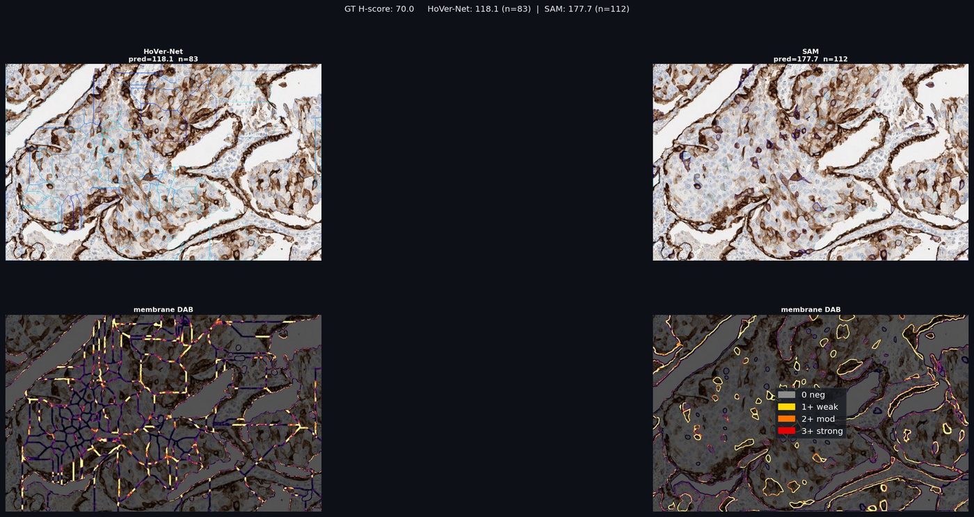
TROP2 IHC per-cell membrane scoring and DAB optical-density analysis

Quantitative TROP2 IHC: per-cell membrane 0/1+/2+/3+ calls, H-score, and DAB optical-density maps -- the reproducible readout that feeds a sharper companion diagnostic.

PV GeoSpatial Intelligence

Local & Community Owned & Operated AI

A BCMG initiative applying production-grade AI -- geospatial, biospatial, and general computer vision -- to problems with direct local and community benefit. Developed with a BCMG ML Engineer. The common thread: rigorous methods, community-scale problems, results that belong to the people they affect.

What We Build

PV GeoSpatial Intelligence draws from the full BCMG platform -- InSAR ground displacement analysis, satellite computer vision, foundation model pathology, and whatever else the problem demands. No single-domain constraint.

Some projects are pro bono where data access, participant engagement, and public benefit align. Others carry commercial licensing: Movement Index data delivered to real estate operators, oil and gas producers, and the City of RPV for community planning and predictive landslide management.

Active area of interest: layering BCMG InSAR displacement history with El Niño precipitation forecasts for anticipatory landslide movement modeling -- early warning at the parcel level, ahead of seasonal acceleration events.

Collaboration Model

Open to academic departments, city agencies, NGOs, and community organizations with defined problems and access to relevant data. Bring the domain knowledge and the data; we bring the computational platform.

Current priorities: South Bay geophysical risk, community health monitoring, and environmental computer vision applications accessible to public-interest stakeholders.

All outputs -- risk scores, maps, classifiers -- remain owned by the community partners where public benefit is the primary use case.


Active · South Bay
PV Movement Index

Per-parcel 0–100 ground displacement score from ALOS-2 InSAR. Covering the Portuguese Bend landslide complex. Commercial licensing to real estate, insurance, and the City of RPV.

Research · Active Interest
Anticipatory Landslide Modeling

BCMG InSAR displacement history + El Niño precipitation forecasts. Parcel-level early warning ahead of seasonal acceleration. Active research interest.

Open · Seeking Partners
Community Health & Environmental CV

Environmental computer vision and community health monitoring. Open to city agencies, NGOs, and academic collaborators with relevant data and community mandate.

Research Materials

BCMG Research Tissues

Over 40,000 FFPE tissue blocks from independent pathology practice, plus breast and prostate tumor/non-tumor tissue microarrays. Blocks and TMAs ship with a digital H&E whole slide image.

Available: Breast Tissue Cohort

200 FFPE blocks across six diagnostic categories. De-identified archival material. WSI scan (SVS) delivered within five business days. Custom selection by diagnosis, grade, or receptor status. Institutional pricing for 20+ blocks.

DCISLCISIDC ILCFibroadenomaBenign FCC
FFPE tissue block$150
WSI scan included
IHC — HER2$100 / block
ER, PR, Ki-67, p53 at reduced rates. Panel pricing on inquiry.
Molecular sequencingOn request
Platform and panel dependent.

Inquire about availability and cohort composition. We respond within 48 hours.


Tissue Microarrays

Breast & Prostate TMAs

Tumor and non-tumor Tissue MicroArrays for breast and prostate, built for high-throughput biomarker and antibody studies. Other organs available on request. Each TMA block ships with a digital H&E whole slide image, and an optional ViewsML virtual biomarker profile for in-silico marker screening before you stain.

Breast · tumorBreast · non-tumor Prostate · tumorProstate · non-tumor Other organs on request
TMA block$1,500
Digital H&E WSI included. Optional ViewsML virtual biomarker profile.
ViewsML virtual biomarker profile: an optional in-silico readout delivered with a TMA -- predicted biomarker profiles from the H&E WSI, so you can prioritize cores and panels before committing stain.

Sourcing Partner

Biobase.ai

Tissue sourcing and biobanking partner. Explore broader cohort availability and custom acquisition through Biobase.

Visit Biobase.ai →

Biobase WSI supplier archive and market demo.

Contract Research

BCMG as a MicroCRO

A small, senior, pathologist-run contract research organization. BCMG delivers routine histology, IHC, and multiplex immunofluorescence (mIF), plus end-to-end project development, in CAP/CLIA-accredited laboratories -- paired with state-of-the-art digital pathology and AI. The advantage of small: a board-certified pathologist is on every project, not three layers away.

Routine Histology
Sectioning, H&E, special stains, and WSI scanning with QC.
Immunohistochemistry
Single and panel IHC with quantitative DAB scoring (H-score, membrane calls) and pathologist review.
Multiplex Immunofluorescence
Opal / Vectra Polaris mIF up to 8-plex, with spectral unmixing and spatial phenotyping.
Virtual H&E from mIF
Physics-based Beer-Lambert pseudo-H&E from one multiplex acquisition -- morphology without cutting an extra section.
Digital Pathology & AI
WSI pipelines, ABMIL classifiers, IHC quantification, cell segmentation, and cloud-native viewing (CloudView).
Project Development
Study design, cohort curation, pipeline build, analysis, and versioned pathologist-signed reporting.

Digital and computational, end to end

BCMG's lab work does not stop at the coverslip. Every specimen can carry through to quantitative image analysis: anchored IHC scoring, mIF spatial phenotyping, cross-modality registration (for example, recovering a CD68 IHC channel into an mIF grid), and scanner color normalization for reproducible cross-site readouts.

Deliverables are the artifacts a sponsor can actually use: QuPath-ready pyramidal images, CSV/JSON feature exports, curated manifests, and self-contained HTML reports signed off by a pathologist.

Virtual H and E rendered from an 8-channel multiplex immunofluorescence whole slide image

Virtual H&E rendered from an 8-channel mIF slide by a physics-based optical-density model -- morphology and immune context from a single acquisition.

CD68 IHC recovered into the multiplex immunofluorescence coordinate grid

Cross-modality salvage: a brightfield CD68 IHC section registered into the mIF grid to recover a macrophage channel.

Services

Consultation Services

Computational pathology and spatial AI expertise for research teams, clinical labs, and industry partners. End-to-end from pipeline design through deployment, licensing, and regulatory guidance.

Computational Pathology
WSI analysis, biomarker quantification, cohort curation, slide QC
Whole Slide Imaging
Scan protocols, tiling pipelines, viewer deployment, DICOM/LIS integration
AI Algorithmic Development & Licensing
ABMIL classifiers, IHC quantification, foundation model benchmarking, algorithm licensing
Multimodal AI — RadPath
Joint radiology-pathology models, cross-modal feature fusion
Computational Pathology Kit (CPK)
BCMG pipeline packaged for on-premises deployment at academic medical centers
CAP/CLIA Laboratory Accreditation & Compliance Guidance
Pathologist-directed guidance for CAP inspection preparation, lab accreditation, and CLIA compliance strategy
IMS / LIS Development
Image management and laboratory information system connectivity
Geospatial Intelligence
InSAR analysis, Movement Index scoring, conflict-zone terrain analysis
IP & Regulatory Strategy
Patent prep, licensing strategy, HIPAA posture review

Describe your project. We respond within 48 hours.

Working with BCMG

  • Board-certified anatomic and clinical pathologist (ABP, 2019)
  • Residency and Urologic Pathology Fellowship, UCLA Medical Center
  • Fellow of the College of American Pathologists (FCAP)
  • Medical degree, Kansas City University of Medicine and Biosciences
  • Published author — peer-reviewed journals + Springer Nature (2022)
  • Active medical licenses: California, Texas
  • S-Corp entity — invoicing and contract-ready
Confidentiality

Non-Disclosure Agreement

Complete and sign the BCMG mutual NDA. Your details populate the agreement in real time. BCMG will countersign and return within 48 hours.

Your Details

Signature

Draw your signature above

Prefer to sign offline? Download template

MUTUAL NON-DISCLOSURE AGREEMENT

Entered into as of , between:

Disclosing Party: Brian Cone Medical Group (S-Corp), Brian Cone, President ("BCMG")

Receiving Party: [Full Name], [Title], [Company]


1. Purpose. The parties wish to explore a business relationship involving BCMG AI tools and IP.

2. Confidential Information. All non-public technical, business, financial, and strategic disclosures are confidential under this Agreement.

3. Obligations. Strict confidence; Purpose use only; no third-party disclosure without consent.

4. Term. Two (2) years from execution.

5. Governing Law. California, County of Los Angeles.


BCMG: (countersignature to follow)

Recipient: draw signature on left

About

Dr. Brian Cone

New Study
BCMG AI x USC -- Clinical-Grade Display Study Underway
A newly started study comparing clinical-grade Barco diagnostic monitors for digital pathology at USC. Dr. William Dean Wallace (USC Keck) is lead investigator; Dr. Cone is co-investigator. Slides supported by a Hamamatsu NanoZoomer scanner.
USC Keck Barco Hamamatsu
Dr. Brian D. Cone, Computational Pathologist

Brian D. Cone, D.O. is a board-certified anatomic and clinical pathologist (American Board of Pathology, 2019) and the founder of Brian Cone Medical Group (BCMG), an independent computational pathology and spatial AI practice based in Southern California.

Dr. Cone completed his residency in Anatomic and Clinical Pathology and a Urologic Pathology Fellowship at UCLA Medical Center, and holds his medical degree from Kansas City University. He is a Fellow of the College of American Pathologists and holds active medical licenses in California and Texas.

His research and practice focus on digital pathology, whole slide image analysis, AI/ML-driven biomarker quantification, geospatial machine learning, and computational tool development. Published author across peer-reviewed journals and a contributing author to Practical Lung Pathology (Springer Nature, 2022). All research, software, and tools presented here are original, independent works conducted through BCMG.

Independence Notice: The products, research, tools, and opinions on this site are solely those of Brian Cone Medical Group (BCMG) and Dr. Cone in his independent consulting capacity. Entirely unrelated to and independent from any current or former employment or contracting engagements. All intellectual property is original BCMG-derived work.

Collaborations

Partners & External Collaborators

Dr. William Dean Wallace
Professor of Pathology, USC Keck School of Medicine. Collaborator on the Pulmonary Hypertension H&E project and joint venture partner on the Computational Pathology Kit (CPK).
BCMG ML Engineering
In-house applied machine learning and data science supporting the BCMG platform across computational pathology, geospatial analysis, and general computer vision.